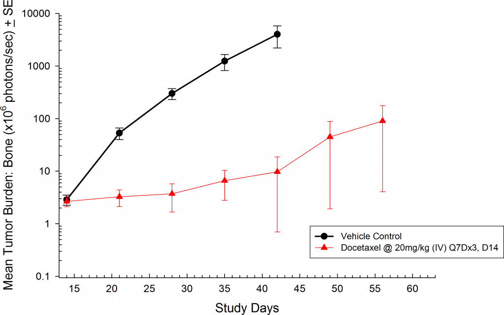
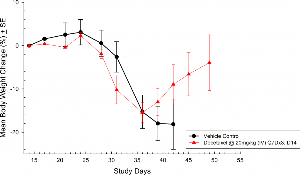
Early detection of prostate cancer is very challenging. Unfortunately patients are asymptomatic until advanced stage disease, leaving them with limited treatment options. The delayed detection also results in an increase in the incidence of metastatic disease. Advanced stage prostate cancer typically metastasizes to the bone and lymph nodes.
Given the poor prognosis (28 percent five-year survival rate) for patients with advanced stage disease (stage III or IV), predictive preclinical models are needed for reliably evaluating treatment options. In most cases of advanced prostate cancer surgical resection is the optimal treatment plan. However, this leaves treating the metastatic lesions with either chemotherapy, radiation, and/or hormone therapy. While subcutaneous xenograft models are valuable, they fail to capture the complexity of the cancer growing in the tissue of origin or the challenges of treating advanced metastatic disease. Orthotopic and metastatic models provide an avenue for not only evaluating the treatment response, but also the impact on the origin tissue and tissue microenvironment, which can be critically important in trying to preserve quality of life.
Orthotopic Modeling
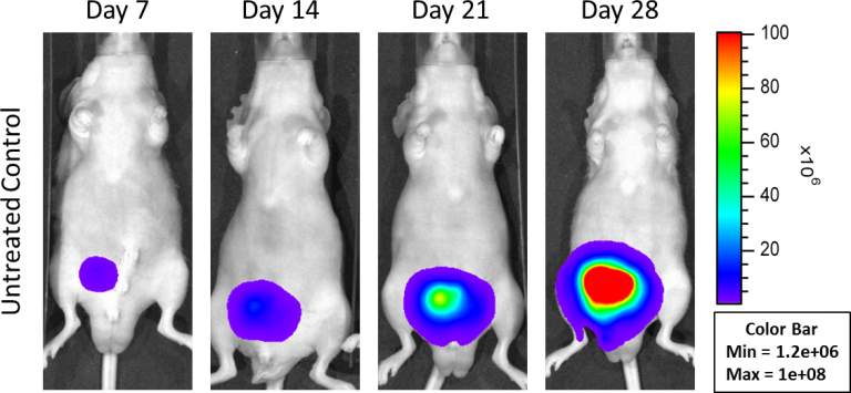
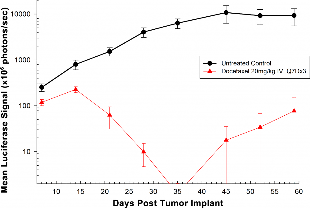
In an effort to address this unmet need, Labcorp has developed an orthotopic and a metastatic prostate model, PC-3M-Luc-C6. This cell line has been transfected with luciferase, which allows for disease and response monitoring through bioluminescence imaging (BLI). The orthotopic model is implanted directly into the prostate gland. Orthotopic tumor growth in the prostate is robust; with established disease present by Day 7 (Figure 1).


